

Description
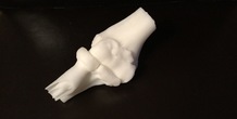
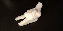
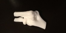
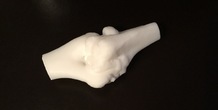
This model of an arthritic elbow was created by a medical student studying a patient's elbow anatomy for a surgical case. The model was printed from the patient specific MRI DICOM files. The surgery the student was involved in was an arthroscopic debridement of osteophytes from the elbow. Arthroscopy uses a camera and instruments inserted into the joint to perform various tasks. An understanding of 3D anatomy is extremely important to prevent getting disoriented and to accomplish the surgical task. The bony arthritic outgrowths in this model are painful and limit motion. Creating a model like this greatly helps students and surgeons-in-training prepare for a wide variety of orthopedic cases. These cases involve topics such as fracture fixation management, joint instability, tumor defect and bone loss repair, deformity alignment correction, and patient specific guides for surgery. The Missouri Orthopedic Institute program allows students the ability to learn the principals of 3D modeling and printing. They develop the skills to independently 3D print medical models for medical education and surgical planning to better educate and care for a diverse patient population.
More from this category
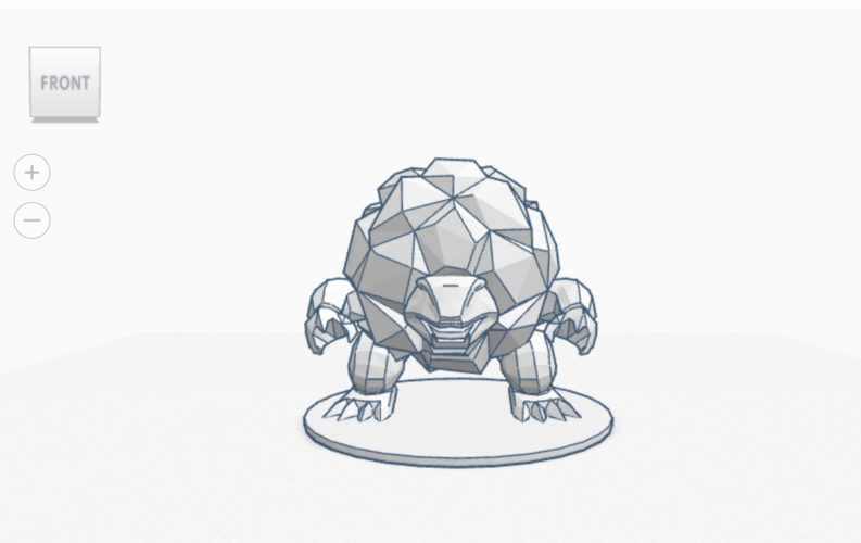

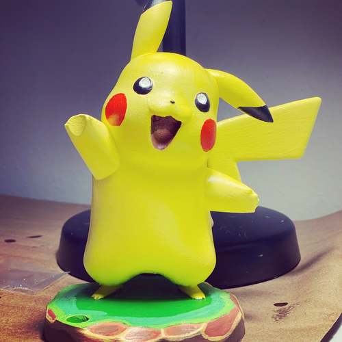
Diglett
by MattSnypes
This is my first stab at TInkerCad. I'll upload a photo as soon as I get a chance to print it out.
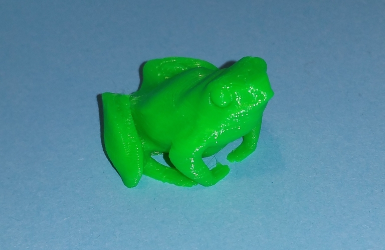
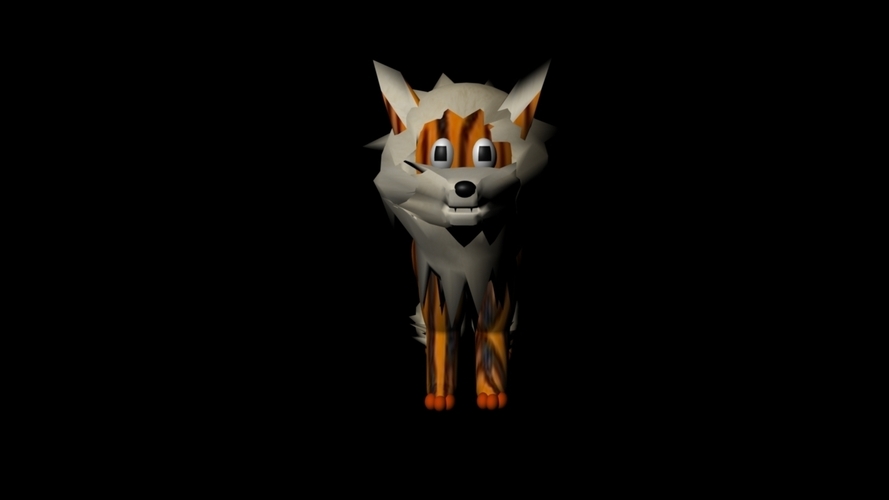
a little frog
by ogolum
I creat this frog, because i love frogs. I scaled it 15x, Print Settings: 0.2mm, no raft, no Supp...

Comments (2)
Sign in to leave a comment.
The Medical Printing Program is revolutionizing healthcare by providing precise and personalized medical documents. This innovative program ensures that patients receive clear and accurate information tailored to their specific needs. For example, patients prescribed <a href="https://borderfreehealth.com/shop/tasigna/">tasigna medication</a> benefit greatly from detailed dosage instructions and potential side effect information provided through this program. By integrating advanced printing technology, the program enhances patient understanding and compliance, ultimately leading to better health outcomes. The Medical Printing Program stands out as a critical tool in modern medical care, bridging the gap between healthcare providers and patients effectively.